Taibai 太白
In the dip just behind (proximal to) the first metatarsophalangeal joint, on the inner side of the foot.
Open the 3D Acupoint Map →
🖼See it in pictures


Location and structures come from our 3D model; only the drawing style was refined with AI. Labels are added by us.

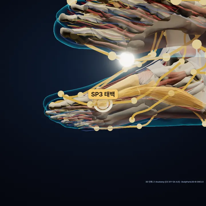
Abductor hallucis · Medial head of flexor hallucis brevis · Sesamoid bones of foot · First metatarsal bone
Rendered straight from the 3D model at the computed location. The glowing ring marks Taibai.
On this page
1Where is Taibai?
In the dip just behind (proximal to) the first metatarsophalangeal joint, on the inner side of the foot.
It sits on the red-white flesh border.
Abductor hallucis · Medial head of flexor hallucis brevis · Sesamoid bones of foot · First metatarsal bone
2How do I find it on my body?
- Find the knobbly joint on the inner side at the base of the big toe.
- Slide just behind it, toward the heel.
- Press the dip there.
- medial head of flexor hallucis brevis
- The inner head of the short big-toe flexor in the sole. It attaches near the inner sesamoid bone under the root of the big toe.
- metatarsophalangeal joint
- The joint where a metatarsal meets the first bone of a toe, the root joint of the toe. Bend the toes and you feel it as a round knuckle on the top of the foot.
- sesamoid bones of foot
- Small seed-shaped bones (usually two) inside the tendons under the root of the big toe. They act as pads that protect the tendons when we step.
- abductor hallucis
- A muscle along the inner edge of the sole from the heel to the big toe. It spreads the big toe and supports the inner arch.
- red-white flesh border
- The border on the hands and feet where the skin of the back meets the skin of the palm or sole and its color and texture change; the old name means 'border of red and white flesh'. It is used as a guide line for locating points.
3Before you press
- Avoid broken or swollen skin and press only to a comfortable level.
- Acupressure is not a substitute for medical care.
4How has it been used traditionally?
- Classified as both the Yuan-source point and the Shu-stream (Earth) point of the Spleen channel (Lingshu, ch. 1 and 2) — a historical classification, not a claim of effect.
- Taibai (太白) was also an old name for the planet Venus.
From traditional texts — not a claim of effect.
5Classical text
Donguibogam (Heo Jun, 1613), Acupuncture and Moxibustion, "Taibai"
在足大指內側核骨下陷中足太陰脈之所注爲腧 …《銅人》
① On the inner side of the big toe, in the hollow below the "kernel bone". Where the Foot Taiyin channel pours in, which makes it the Shu-stream point (Tongren).
- Source tags as given in the Donguibogam: Tongren = Tongren Shuxue Zhenjiu Tujing (1026).
- The garbled glyph 杮 has been corrected to 腧, checked against Lingshu ch.
- 2 and the Zhenjiu Jiayi Jing vol.
- 3.
- The "kernel bone" (hegu) is an old name for the rounded bony bump at the inner base of the big toe.
- "…" marks the needling depth and moxa count, left out because they are for licensed practitioners.
Original text as written · translation is ours.
?FAQ
Where exactly is Taibai?
In the dip just behind (proximal to) the first metatarsophalangeal joint, on the inner side of the foot. It sits on the red-white flesh border.
How do I find Taibai?
Find the knobbly joint on the inner side at the base of the big toe. Slide just behind it, toward the heel. Press the dip there. you are on the line where top-of-foot skin meets sole skin.
What should I watch out for?
Avoid broken or swollen skin and press only to a comfortable level. Acupressure is not a substitute for medical care.
Sources
- Location: WHO Standard Acupuncture Point Locations in the Western Pacific Region (2008), p. 71
- Standard comparison: Chinese national standard GB/T 12346-2021 (Nomenclature and location of meridian points)
- Korean anatomical terms and locating tips: KMCRIC Standard Acupoint DB (Korea)
- Classification source: Lingshu ch. 1 (Jiu Zhen Shi Er Yuan, source point) and ch. 2 (Ben Shu); Nanjing ch. 64 for the phases
- Traditional use: Hyeoljari Seodang (Bookdramang, 2015; Korean), ch. 4 "Taibai" and appendix (Yixue Rumen indications)
- Classical text: Donguibogam, Acupuncture and Moxibustion, "Taibai" — Wikisource public edition (garbled glyph 杮 corrected to 腧)
- Indications: Yixue Rumen vol. 1 (Wikisource)
- 3D: computed on Z-Anatomy (CC BY-SA 4.0) · BodyParts3D © DBCLS
Positions computed from the WHO standard locations (2008) on Z-Anatomy (CC BY-SA 4.0) · BodyParts3D © DBCLS. For reference only — not a substitute for diagnosis or treatment. See a clinician if you are unwell.