Neiting 內庭
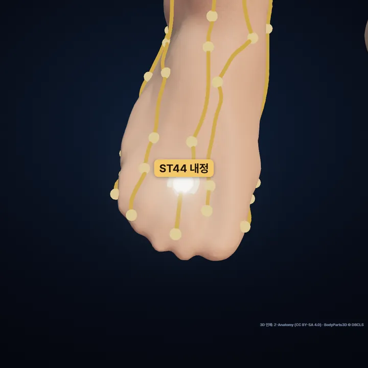
Between the second and third toes, just behind the edge of the web.
Open the 3D Acupoint Map →
🖼See it in pictures
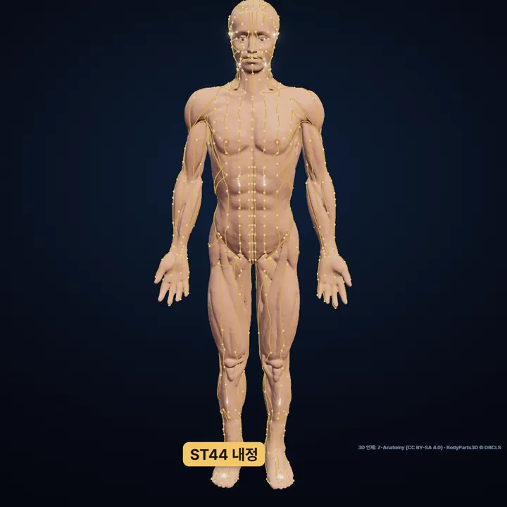

Location and structures come from our 3D model; only the drawing style was refined with AI. Labels are added by us.
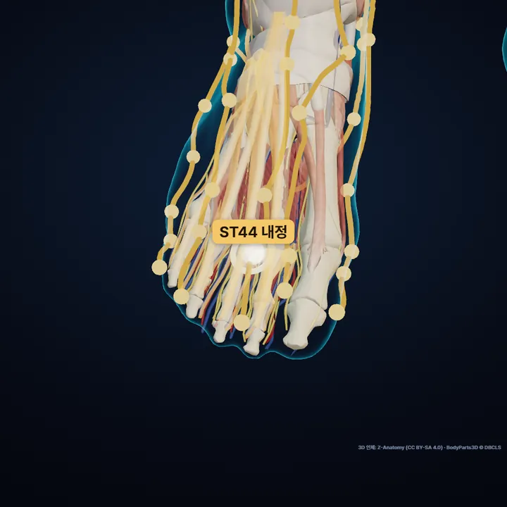
Proximal phalanx of second finger of foot · Dorsal digital arteries of foot · Tendon of extensor digitorum longus · Extensor digitorum brevis
Rendered straight from the 3D model at the computed location. The glowing ring marks Neiting.
On this page
1Where is Neiting?
Between the second and third toes, just behind the edge of the web.
It sits on the red-white flesh border, where top-of-foot skin meets sole skin.
Proximal phalanx of second finger of foot · Dorsal digital arteries of foot · Tendon of extensor digitorum longus · Extensor digitorum brevis
2How do I find it on my body?
- Spread the second and third toes slightly and find the web between them.
- Move a little back from the web's edge toward the top of the foot.
- tendon of extensor digitorum longus
- The four strands the extensor digitorum longus splits into on the top of the foot. Lift your toes and they stand out like cords.
- dorsal digital arteries of foot
- The thin arteries running to the toes on the top of the foot.
- extensor digitorum brevis
- A flat muscle on the outer top of the foot. Lift your toes and it bulges as a small soft lump in front of and below the outer ankle.
- red-white flesh border
- The border on the hands and feet where the skin of the back meets the skin of the palm or sole and its color and texture change; the old name means 'border of red and white flesh'. It is used as a guide line for locating points.
- proximal phalanx
- The first bone of a finger or toe, at the root end. It forms a joint with the metacarpal or metatarsal bone.
3Before you press
- Avoid broken or swollen skin and press only to a comfortable level.
- Acupressure is not a substitute for medical care.
4How has it been used traditionally?
- Classified as the Ying-spring (Water) point of the Stomach channel — a historical classification, not a claim of effect.
- The name joins nei (inner) and ting (courtyard): a small yard tucked inside the fork of the toes.
From traditional texts — not a claim of effect.
5Classical text
Donguibogam (Heo Jun, 1613), Acupuncture and Moxibustion, "Neiting"
在足大指次指外間陷中《銅人》
在足次指與三指岐骨間陷中《入門》
足陽明脈之所流爲滎 …《銅人》
- ①In the hollow in the gap on the outer side of the second toe. Tongren
- ②In the hollow where the bones of the second and third toes fork. Rumen
- ③Where the Foot Yangming channel trickles on, which makes it the Ying-spring point. Tongren
- Source tags as given in the Donguibogam: Tongren = Tongren Shuxue Zhenjiu Tujing (1026); Rumen = Yixue Rumen (1575).
- This entry has no garbled glyphs.
- "…" marks the needling depth and moxa count, left out because they are for licensed practitioners.
Original text as written · translation is ours.
?FAQ
Where exactly is Neiting?
Between the second and third toes, just behind the edge of the web. It sits on the red-white flesh border, where top-of-foot skin meets sole skin.
How do I find Neiting?
Spread the second and third toes slightly and find the web between them. Move a little back from the web's edge toward the top of the foot. you are on the boundary where the darker skin of the top meets the paler skin of the sole.
What should I watch out for?
Avoid broken or swollen skin and press only to a comfortable level. Acupressure is not a substitute for medical care.
Sources
- Location: WHO Standard Acupuncture Point Locations in the Western Pacific Region (2008), p. 68
- Standard comparison: Chinese national standard GB/T 12346-2021 (Nomenclature and location of meridian points)
- Korean anatomical terms and locating tips: KMCRIC Standard Acupoint DB (Korea)
- Classification source: Lingshu ch. 2 (Ben Shu) for the five transport points; Nanjing ch. 64 for the phases; Zhenjiu Jiayi Jing vol. 3 (Siku Quanshu edition)
- Traditional use: Hyeoljari Seodang (Bookdramang, 2015; Korean), ch. 3 "Neiting" and appendix (Yixue Rumen indications)
- Classical text: Donguibogam, Acupuncture and Moxibustion, "Neiting" — Wikisource public edition (no garbled glyphs in this entry)
- Indications: Yixue Rumen vol. 1 (Wikisource)
- 3D: computed on Z-Anatomy (CC BY-SA 4.0) · BodyParts3D © DBCLS
Positions computed from the WHO standard locations (2008) on Z-Anatomy (CC BY-SA 4.0) · BodyParts3D © DBCLS. For reference only — not a substitute for diagnosis or treatment. See a clinician if you are unwell.