Lidui 厲兌
0.1 F-cun back and out from the outer corner of the second toenail.
Open the 3D Acupoint Map →
🖼See it in pictures
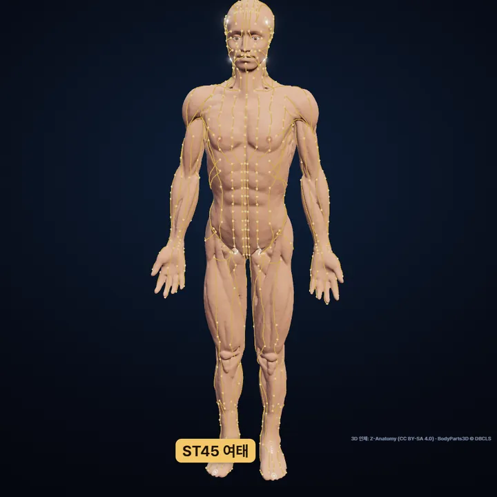
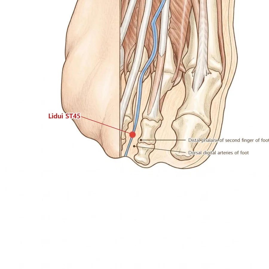
Location and structures come from our 3D model; only the drawing style was refined with AI. Labels are added by us.
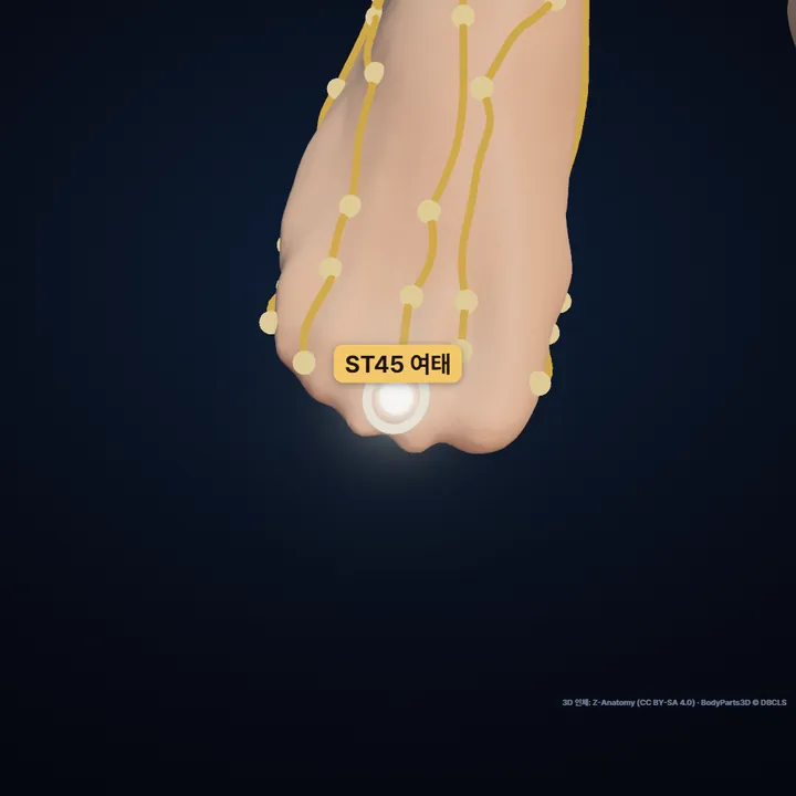
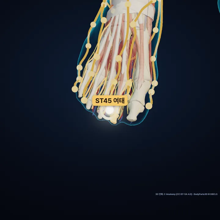
Distal phalanx of second finger of foot · Dorsal digital arteries of foot
Rendered straight from the 3D model at the computed location. The glowing ring marks Lidui.
On this page
1Where is Lidui?
0.1 F-cun back and out from the outer corner of the second toenail.
On the outer side of the end bone, it is where a line along the nail's outer edge meets a line across the nail's base.
Distal phalanx of second finger of foot · Dorsal digital arteries of foot
2How do I find it on my body?
- Picture a line along the outer (little-toe side) edge of the second toenail.
- Picture a line across the base of the nail, where it emerges from the skin.
- Press where the two lines cross.

A cun is a body-scaled unit of length. The precise way is to divide a bone length into equal parts (proportional bone cun); finger widths are a quick estimate.
- dorsal digital arteries of foot
- The thin arteries running to the toes on the top of the foot.
- distal phalanx
- The last bone at the tip of a finger or toe. The nail lies on top of it.
3Before you press
- Skip this point if the skin around the toenail is infected or injured.
- Avoid broken or swollen skin and press only to a comfortable level.
- Acupressure is not a substitute for medical care.
4How has it been used traditionally?
- Classified as the Jing-well (Metal) point of the Stomach channel, where the channel ends — a historical classification, not a claim of effect.
- Classical texts place it a leek leaf's width from the corner of the second toenail.
From traditional texts — not a claim of effect.
5Classical text
Donguibogam (Heo Jun, 1613), Acupuncture and Moxibustion, "Lidui"
在足大指次指端外側去爪甲如韭葉足陽明脈之所出爲井 …《銅人》
① On the outer side of the tip of the second toe, a leek leaf's width from the nail. Where the Foot Yangming channel emerges, which makes it the Jing-well point (Tongren).
- Source tags as given in the Donguibogam: Tongren = Tongren Shuxue Zhenjiu Tujing (1026).
- Garbled glyphs corrected: 崲 to 厲 (the point name) and 堛 to 韭 (leek), checked against Lingshu ch.
- 2 and the Zhenjiu Jiayi Jing vol.
- 3.
- Lingshu ch.
- 2 words the side differently from the Donguibogam; our location follows WHO (lateral).
- "…" marks the needling depth and moxa count, left out because they are for licensed practitioners.
Original text as written · translation is ours.
?FAQ
Where exactly is Lidui?
0.1 F-cun back and out from the outer corner of the second toenail. On the outer side of the end bone, it is where a line along the nail's outer edge meets a line across the nail's base.
How do I find Lidui?
Picture a line along the outer (little-toe side) edge of the second toenail. Picture a line across the base of the nail, where it emerges from the skin. Press where the two lines cross. about 0.1 F-cun diagonally from the nail corner.
What should I watch out for?
Skip this point if the skin around the toenail is infected or injured. Avoid broken or swollen skin and press only to a comfortable level. Acupressure is not a substitute for medical care.
Sources
- Location: WHO Standard Acupuncture Point Locations in the Western Pacific Region (2008), p. 68
- Standard comparison: Chinese national standard GB/T 12346-2021 (Nomenclature and location of meridian points)
- Korean anatomical terms and locating tips: KMCRIC Standard Acupoint DB (Korea)
- Classification source: Lingshu ch. 2 (Ben Shu) for the five transport points; Nanjing ch. 64 for the phases; Zhenjiu Jiayi Jing vol. 3 (Siku Quanshu edition)
- Traditional use: Hyeoljari Seodang (Bookdramang, 2015; Korean), ch. 3 "Lidui" and appendix (Yixue Rumen indications)
- Classical text: Donguibogam, Acupuncture and Moxibustion, "Lidui" — Wikisource public edition (garbled glyphs 崲 corrected to 厲 and 堛 to 韭)
- Indications: Yixue Rumen vol. 1 (Wikisource)
- 3D: computed on Z-Anatomy (CC BY-SA 4.0) · BodyParts3D © DBCLS
Positions computed from the WHO standard locations (2008) on Z-Anatomy (CC BY-SA 4.0) · BodyParts3D © DBCLS. For reference only — not a substitute for diagnosis or treatment. See a clinician if you are unwell.